Showing posts with label Medical Matters. Show all posts

4/27/2013

Tests on Hereditary Diseases

Scientists at the University of Medicine and Dentistry of New Jersey-New Jersey Medical School (UMDNJ-NJMS) have developed new DNA sequencing tests that hold significant promise for decreasing costs associated with diagnosing cancer and hereditary diseases, including cystic fibrosis.

According to the Cystic Fibrosis Foundation web site, "More than 10 million Americans are symptomless carriers of the defective CF gene." This chronic disease impacts the lungs and the digestive system. It occurs when a child inherits one defective CF gene from each parent. Statistics show New Jersey averages 125,000 births of children who are diagnosed with cystic fibrosis annually.Officials at the New Jersey Department of Health approved the use of the new Cystic Fibrosis (CF) Carrier and Diagnosis Test, which was created at the Institute for Genomic Medicine at UMDNJ-NJMS. Using a semiconductor mechanism that was developed by San Francisco-based Ion Torrent, the microchip tests the entire gene for mutations. IGM now offers this certified Clinical Diagnostic Laboratory service for hospitals as well as obstetrics and gynecology practices throughout the Garden State.
"We believe the adaptation of this new sequencing technology will drastically improve our ability to analyze genetic disorders," said Marvin N. Schwalb, PhD, director of the Institute for Genomic Medicine. "Traditional CF sequencing testing costs thousands of dollars making the test unavailable for carrier screening. This new test costs less than $200. Most importantly, the genetic carrier test we developed improves the diagnosis rate to 98 percent. While the test provides significant improvement for all populations, the improved rate is particularly valuable for minorities because current carrier screening methods only detects approximately 65% of mutations in these populations."
The new technology provides many advances including the ability to test as many as 96 samples on a single platform and the fact that the equipment cost 1/10 as much as the previous technology.
IGM has developed another test, which was also approved by the NJHSS, for mitochondrial DNA. Mutations in mitochondria cause a wide variety of diseases, such eye and neuromuscular system disorders and possible cancer.
Schwalb, a professor of Pediatrics, Microbiology and Molecular Genetics at NJMS, said, "We are proud of the fact that the IGM is a world leader in the advancement of genetic diagnosis. DNA sequencing will keep us very busy for a while. In the state of New Jersey, there is nothing that compares to this advancement and this is just the beginning."
Source: University of Medicine and Dentistry of New Jersey (UMDNJ) (2012, August 16). Researchers develop DNA sequencing tests for hereditary diseases. ScienceDaily. Retrieved April 27, 2013, from http://www.sciencedaily.com/releases/2012/08/120816170309.htm?utm_source=feedburner&utm_medium=feed&utm_campaign=Feed%3A+sciencedaily+%28ScienceDaily%3A+Latest+Science+News%29

4/25/2013

Gut bacteria linked to obesity

Researchers at the University of Maryland School of Medicine have identified 26 species of bacteria in the human gut microbiota that appear to be linked to obesity and related metabolic complications. These include insulin resistance, high blood sugar levels, increased blood pressure and high cholesterol, known collectively as "the metabolic syndrome," which significantly increases an individual’s risk of developing diabetes, cardiovascular disease and stroke.

"We identified 26 species of bacteria that were correlated with obesity and metabolic syndrome traits such as body mass index (BMI), triglycerides, cholesterol, glucose levels and C-reactive protein, a marker for inflammation," says the senior author, Claire M. Fraser, Ph.D., professor of medicine and microbiology and immunology and director of the Institute for Genome Sciences (IGS) at the University of Maryland School of Medicine. "We can’t infer cause and effect, but it’s an important step forward that we're starting to identify bacteria that are correlated with clinical parameters, which suggests that the gut microbiota could one day be targeted with medication, diet or lifestyle changes."The results of the study, which analyzed data from the Old Order Amish in Lancaster County, Pa., are being published online on Aug. 15, 2012, in PLOS ONE, which is published by the Public Library of Science (PLOS One). The study was funded by the National Institutes of Health (NIH). (UH2/UH3 DK083982, U01 GM074518 and P30 DK072488)
Dr. Fraser says that additional research, including an interventional study with the Amish, is essential. "We can look at whether these bacteria change over time in a given individual or in response to diet or medication," she says.
Dr. Fraser notes that the research team, led by Margaret L. Zupancic, Ph.D., then a postdoctoral fellow at IGS, also found an apparent link between the gut bacteria and inflammation, which is believed to be a factor in obesity and many other chronic diseases. "This is one of the first studies of obesity in humans to make a link between inflammatory processes and specific organisms that are present in the GI tract," Dr. Fraser says, noting that participants with metabolic syndrome who had elevated serum markers associated with inflammation tended to have the lowest levels of good bacteria that have been reported previously to have anti-inflammatory properties.
The study is the result of an ongoing collaboration between Dr. Fraser and Alan R. Shuldiner, M.D., in connection with the NIH’s Human Microbiome Project, which seeks to characterize microbial communities in the body. Dr. Shuldiner, associate dean for personalized medicine and director of the Program in Personalized and Genomic Medicine at the University of Maryland School of Medicine, operates an Amish research clinic in Lancaster Pa. Over the past 20 years, he and his research team have conducted more than a dozen studies with the Amish, looking for genes that may cause common diseases, such as diabetes, osteoporosis and cardiovascular disease.
"The Old Order Amish are ideal for such studies because they are a genetically homogenous population descended from a few founder families and have a similar rural lifestyle," Dr. Shuldiner, the John L. Whitehurst Professor of Medicine, says. "We believe the results of this study are relevant to a broader population because the clinical characteristics of obesity and its complications in the Amish are no different from the general Caucasian population," he says.
E. Albert Reece, M.D., Ph.D., M.B.A., vice president for medical affairs at the University of Maryland and the John Z. and Akiko K. Bowers Distinguished Professor and dean of the University of Maryland School of Medicine, says, "Obesity and its related complications have become a critical public health concern, and the number of people who are now considered obese or overweight has skyrocketed. Dr. Fraser and Dr. Shuldiner are two of our most senior research-scientists and leaders in their respective fields. This study provides valuable insights into the role the bacteria in our bodies may play in obesity and the metabolic syndrome. We may ultimately be able to target the gut microbiome to help prevent or mitigate risk factors for a number of diseases."
The researchers analyzed the bacteria in fecal samples of 310 members of the Old Order Amish community, using a process that enables them to identify a marker gene that serves as a bar code for each type of bacteria. Participants in the study ranged from lean to overweight to obese; some of the obese participants also had features of the metabolic syndrome. "Our hypothesis was that we would see a different composition in the gut microbiota in lean vs. obese individuals and possibly in individuals who were obese but also had features of the metabolic syndrome."
They discovered that every individual possessed one of three different communities of interacting bacteria, each characterized by a dominant bacterial genus. Neither BMI nor any metabolic syndrome trait was specifically associated with any of these communities. Instead, differing levels of 26 less abundant bacterial species present in all individuals appeared to be linked to obesity and certain features of the metabolic syndrome.
Interestingly, researchers also analyzed people's gut bacteria by their occupation and found that those who had regular contact with livestock, such as farmers and their wives, had bacterial communities dominated by Prevotella, a type of bacteria that is also abundant in the gut microbiota of cattle and sheep. "These findings suggest that environmental exposure may play a role in determining the composition of the gut microbiota in humans," Dr. Fraser says.

University of Maryland Medical Center (2012, August 15). Gut bacteria linked to obesity and metabolic syndrome identified. ScienceDaily. Retrieved April 26, 2013, from http://www.sciencedaily.com­/releases/2012/08/120815174902.htm?utm_source=feedburner&utm_medium=feed&utm_campaign=Feed%3A+sciencedaily+%28ScienceDaily%3A+Latest+Science+News%29

4/24/2013

Scientists Can Now Block Heroin, Morphine Addiction

In a major breakthrough, an international team of scientists has proven that addiction to morphine and heroin can be blocked, while at the same time increasing pain relief.


Laboratory studies have shown that the drug (+)-naloxone (pronounced: PLUS nal-OX-own) will selectively block the immune-addiction response.The team from the University of Adelaide and University of Colorado has discovered the key mechanism in the body's immune system that amplifies addiction to opioid drugs.
The results -- which could eventually lead to new co-formulated drugs that assist patients with severe pain, as well as helping heroin users to kick the habit -- will be published August 16 in the Journal of Neuroscience.
"Our studies have shown conclusively that we can block addiction via the immune system of the brain, without targeting the brain's wiring," says the lead author of the study, Dr Mark Hutchinson, ARC Research Fellow in the University of Adelaide's School of Medical Sciences.
"Both the central nervous system and the immune system play important roles in creating addiction, but our studies have shown we only need to block the immune response in the brain to prevent cravings for opioid drugs."
The team has focused its research efforts on the immune receptor known as Toll-Like receptor 4 (TLR4).
"Opioid drugs such as morphine and heroin bind to TLR4 in a similar way to the normal immune response to bacteria. The problem is that TLR4 then acts as an amplifier for addiction," Dr Hutchinson says.
"The drug (+)-naloxone automatically shuts down the addiction. It shuts down the need to take opioids, it cuts out behaviours associated with addiction, and the neurochemistry in the brain changes -- dopamine, which is the chemical important for providing that sense of 'reward' from the drug, is no longer produced."
Senior author Professor Linda Watkins, from the Center for Neuroscience at the University of Colorado Boulder, says: "This work fundamentally changes what we understand about opioids, reward and addiction. We've suspected for some years that TLR4 may be the key to blocking opioid addiction, but now we have the proof.
"The drug that we've used to block addiction, (+)-naloxone, is a non-opioid mirror image drug that was created by Dr Kenner Rice in the 1970s. We believe this will prove extremely useful as a co-formulated drug with morphine, so that patients who require relief for severe pain will not become addicted but still receive pain relief. This has the potential to lead to major advances in patient and palliative care," Professor Watkins says.
The researchers say clinical trials may be possible within the next 18 months.
This study has been funded by the National Institute on Drug Abuse (NIDA) in the United States and the Australian Research Council (ARC).
Source: University of Adelaide (2012, August 14). Scientists can now block heroin, morphine addiction. ScienceDaily. Retrieved April 24, 2013, from http://www.sciencedaily.com­/releases/2012/08/120814213246.htm

4/15/2013

Half of Inhaled Soot Particles from Diesel Exhaust, Fires Gets Stuck in the Lungs


The exhaust from diesel-fueled vehicles, wood fires and coal-driven power stations contains small particles of soot that flow out into the atmosphere. The soot is a scourge for the climate but also for human health. Now for the first time, researchers have studied in detail how diesel soot gets stuck in the lungs. The results show that more than half of all inhaled soot particles remain in the body. (Credit: © Imagenatural / Fotolia)

The figure is higher than for most other types of particles. For example "only" 20 per cent of another type of particle from wood smoke and other biomass combustion gets stuck in the lungs. One explanation is that diesel soot is made up of smaller particles and can therefore penetrate deeper into the lungs, where it is deposited. The study was made on diesel particles (which mainly consist of soot) and was recently published in the Journal of Aerosol Science. Ten healthy people volunteered for the the study.
"Findings of this kind can be extremely useful both for researchers to determine what doses of soot we get into our lungs out of the amount we are exposed to, and to enable public authorities to establish well-founded limits for soot particles in outdoor air," says Jenny Rissler, researcher in aerosol technology at Lund University's Faculty of Engineering and responsible for publishing the study.
In population studies, other researchers have been able to observe that people who live in areas with high concentrations of particulates are more affected by both respiratory and cardiovascular diseases. But since there is no conclusive evidence that it is precisely the soot that is to blame, the authorities have so far not taken any decisions on guidelines.
"Currently there is no specific limit for soot particles in the air, despite the fact that soot in the air is linked to both lung cancer and other diseases," says Jenny Rissler.
But Jenny Rissler thinks that in the future, limits on soot levels will also be set, with reference to the WHO's recent reclassification of diesel exhaust from "probably carcinogenic" to "carcinogenic."
Soot particles are not only connected to effects on health but may also contribute to a warmer climate. Paradoxically, other types of aerosol particles can partly be desirable, insofar as they have a cooling effect on the climate and thereby mitigate the warming effect of carbon dioxide.
"Soot particles are black and absorbs light, thus producing a warming effect. So it could be a double advantage to reduce it," she observes.
Jenny Rissler will next be studying individual variations in lung deposition and exposing cells to soot. She is also in the process of further developing methods to measure the surface area of the particles, as this has shown to be an important indicator of their harmfulness.
Background: Every time we breathe, we inhale tiny airborne particles, so-called aerosol particles. Some occur naturally, while others are the result of human activity. Soot mainly belongs in the latter category, as a by-product of combustion from power stations to small-scale wood fires and decorative candles. Another common source of soot is the exhaust from diesel engines, even though modern diesel cars have considerably reduced emissions thanks to efficient filters.
The EU will be tightening rules on emissions for heavy duty diesel vehicles in 2014.
-------------
The above story is reprinted from materials provided byLund University.
Jenny Rissler, Erik Swietlicki, Agneta Bengtsson, Christoffer Boman, Joakim Pagels, Thomas Sandström, Anders Blomberg, Jakob Löndahl. Experimental determination of deposition of diesel exhaust particles in the human respiratory tractJournal of Aerosol Science, 2012; 48: 18 DOI: 10.1016/j.jaerosci.2012.01.005
Lund University (2012, June 27). Half of inhaled soot particles from diesel exhaust, fires gets stuck in the lungs. ScienceDaily. Retrieved April 15, 2013, from http://www.sciencedaily.com­/releases/2012/06/120627092016.htm

6/21/2012

Avian Flu Viruses Which Are Transmissible Between Humans Could Evolve in Nature


It might be possible for human-to-human airborne transmissible avian H5N1 influenza viruses to evolve in nature, new research has found.
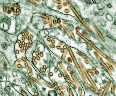
Avian Flu Viruses Which Are Transmissible Between Humans Could Evolve in Nature
Colorized transmission electron micrograph of Avian influenza A H5N1 viruses (seen in gold) grown in MDCK cells (seen in green) [Credit: CDC/Courtesy of Cynthia Goldsmith; Jacqueline Katz; Sherif R. Zaki]
The findings, from research led by Professor Derek Smith and Dr Colin Russell at the University of Cambridge, were published June 22 in the journal Science.

Currently, avian H5N1 influenza, also known as bird flu, can be transmitted from birds to humans, but not (or only very rarely) from human to human. However, two recent papers by Herfst, Fouchier and colleagues in Science and Imai, Kawaoka and colleagues in Nature reveal that potentially with as few as five mutations (amino acid substitutions), or four mutations plus reassortment, avian H5N1 can become airborne transmissible between mammals, and thus potentially among humans. However, until now, it was not known whether these mutations might evolve in nature.

The Cambridge researchers first analysed all of the surveillance data available on avian H5N1 influenza viruses from the last 15 years, focusing on birds and humans. They discovered that two of the five mutations seen in the experimental viruses (from the Fouchier and Kawaoka labs) had occurred in numerous existing avian flu strains. Additionally, they found that a number of the viruses had both of the mutations.

Colin Russell, Royal Society University Research Fellow at the University of Cambridge, said: "Viruses that have two of these mutations are already common in birds, meaning that there are viruses that might have to acquire only three additional mutations in a human to become airborne transmissible. The next key question is 'is three a lot, or a little?' "

The scientists explored this key question using a mathematical model of how viruses replicate and evolve within a mammalian host and assessed the influence of various factors on whether the remaining three mutations could evolve in a single host or in a short chain of transmission between hosts

The factors that increased the likelihood of mutations evolving are:

1. Random mutation. The replication mechanisms of influenza viruses don't make perfect copies. On average, every time an influenza virus replicates itself it makes approximately one mutation somewhere in the genome of each new virus. In each infected human there will be billions of viruses, and thus with many viruses replicating, multiple mutations can accumulate within a single host.

2. Positive selection. If some of the remaining mutations help the avian virus to adapt to mammals, then those mutations will make the viruses more fit and thus will be positively selected and preferentially accumulate.

3. Long infection. The longer someone is infected and producing new viruses, the more time there is for mutations to accumulate.

4. Functionally equivalent substitutions. The sets of substitutions identified by Fouchier and Kawaoka are unlikely to be the only combinations of substitutions capable of producing an aerosol transmissible virus. The probability of emergence increases with the number of combinations.

5. Diversity in the within-bird virus population. Given all of the mutations there are likely to be within a host due to random mutation, it is possible that the viruses from a bird that infect a human might have a mutation that would not be detected by routine surveillance. For example, if 100 virus particles from a bird infect a human and one of those particles had a key mutation, it would increase the probability of the mutation reaching high levels within a host even though routine sequencing would not detect it.

6. Transmission between mammals. If mammals are capable of transmitting viruses that have some but not all of the necessary substitutions it could increase the probability of an airborne transmissible virus evolving.

The factors that decreased the likelihood of mutations evolving are:

1. An effective immune response. An effective immune response would shorten the length of an infection and thus decrease the time available to accumulate mutations.

2. Deleterious substitutions. If any of the substitutions necessary for airborne transmission were harmful to the virus it would, on average, slow the accumulation of mutations.

3. Order of acquiring mutations. It is not currently known if the mutations for airborne transmissibility need to be acquired in a specific order. If they do, it would, on average, slow the accumulation of mutations.

"With the information we have, it is impossible to say what the exact risk is of the virus becoming airborne transmissible among humans. However, the results suggest that the remaining three mutations could evolve in a single human host, making a virus evolving in nature a potentially serious threat," said Derek Smith, Professor of Infectious Disease Informatics at the University of Cambridge. "We now know that it is in the realm of possibility that these viruses can evolve in nature, and what needs to be done to assess the risk more accurately of these mutations evolving in nature."

The scientists recommend the following activities be considered high priority for estimating and ameliorating the risk of emergence of aerosol transmissible H5N1 viruses.

First, additional surveillance in regions where viruses with airborne transmission enabling substitutions have been observed and in regions connected to those regions by bird migration and trade. Also, increased surveillance for mutations that might have the same function as those found by the Fouchier and Kawaoka labs.

Second, related to surveillance, some targeted sequencing of H5N1 viruses should be done by "deep sequencing" where the lab sequences many viruses from an individual host to look for viruses that might have accumulated the critical mutations, even if those viruses are just a small proportion of the viruses within an animal.

Third, further investigations are needed to determine which substitutions and combinations of substitutions that are not the same as, but have the same function as, the substitutions identified by the Fouchier and Kawaoka labs are capable of making viruses airborne transmissible between mammals.

Fourth, further studies are needed to elucidate the changes in within-host fitness and between-host transmissibility associated with each airborne transmission enabling substitution and combination of substitutions.

Professor Smith added: "The situation is similar to assessing the risk of an earthquake or tsunami. We don't know exactly when and where, but by increasing monitoring and research -- some of which is already underway -- scientists and public health officials will be able to increase the accuracy with which the risk can be assessed and to minimise those risks."

The research was funded by multiple sources including the European Commission through framework 7 grants EMPERIE and ANTIGONE, the Royal Society, the Human Frontiers Science Program, the Wellcome Trust, and the National Institutes of Health.

Source: University of Cambridge [June 21, 2012]

6/06/2012

1 Million Billion Billion Billion Billion Billion Billion: Number of Undiscovered Drugs


A new voyage into "chemical space" -- occupied not by stars and planets but substances that could become useful in everyday life -- has concluded that scientists have synthesized barely one tenth of 1 percent of the potential medicines that could be made. The report, in the journal ACS Chemical Neuroscience, estimates that the actual number of these so-called "small molecules" could be 1 novemdecillion (that's 1 with 60 zeroes), 1 million billion billion billion billion billion billion, which is more than some estimates of the number of stars in the universe.

1 Million Billion Billion Billion Billion Billion Billion: Number of Undiscovered Drugs
A new voyage into "chemical space" -- occupied not by stars and planets but substances that could become useful in everyday life -- has concluded that scientists have synthesized barely one tenth of 1 percent of the potential medicines that could be made [Credit: Web]
Jean-Louis Reymond and Mahendra Awale explain that small molecules, which are able to cross cell walls and interact with biological molecules in the body, are prime targets for scientists who develop new medicines. Most existing medications are small molecules. The authors focused on the "chemical space" inhabited by all of the small molecules that could possibly exist according to the laws of physics and chemistry. 

Researchers have identified millions of these compounds -- the ACS' Chemical Abstracts Service database contains almost 67 million substances. Reymond and Awale estimate that the molecules synthesized and tested as potential drugs so far represent less than 0.1 percent of chemical space. To aid researchers looking for new ways to prevent and treat disease, they set out to find the best ways to search for new small molecules.

The authors discuss several ways of getting a handle on chemical space, including by the size, shape and makeup of molecules. They show how computers can help researchers efficiently narrow a search for a new drug candidate. Computer modeling of chemical interactions can help researchers find a handful of promising molecules to synthesize and test in the lab. "Small molecule drugs are essential to the success of modern medicine," the authors note, and suggest that their methods may be particularly useful for finding new pharmaceuticals that target the central nervous system.

Source: American Chemical Society [June 06, 2012]

6/05/2012

Air Pollution Linked to Chronic Heart Disease


Air pollution, a serious danger to the environment, is also a major health risk, associated with respiratory infections, lung cancer and heart disease. Now a Tel Aviv University researcher has concluded that not only does air pollution impact cardiac events such as heart attack and stroke, but it also causes repeated episodes over the long term.

Air Pollution Linked to Chronic Heart Disease

Cardiac patients living in high pollution areas were found to be over 40 percent more likely to have a second heart attack when compared to patients living in low pollution areas, according to Dr. Yariv Gerber of TAU's School of Public Health at the Sackler Faculty of Medicine. "We know that like smoking cigarettes, pollution itself provokes the inflammatory system. If you are talking about long-term exposure and an inflammatory system that is irritated chronically, pollution may well be involved in the progression of atrial sclerosis that manifests in cardiac events," explains Dr. Gerber.

Done in collaboration with Prof. Yaacov Drory and funded by the Environmental and Health Fund in Jerusalem, the research was presented at the San Diego Epidemiological Meeting of the American Heart Association in March and the Annual Meeting of the Israeli Heart Society in April.

Risking recurrence

Air pollution has previously been acknowledged as a factor in heart attack risk, as well as other health risks. The goal of this study, says Dr. Gerber, was to quantify that association and determine the long-term effects of air pollution on myocardial infarction (MI) patients. Their study followed 1,120 first-time MI patients who had been admitted to one of eight hospitals in central Israel between 1992 and 1993, all of whom were under the age of 65 at the time of admittance. The patients were followed up until 2011, a period of 19 years.

Air quality was measured at 21 monitoring stations inareas where the patients lived, and analyzed by a group of researchers at the Technion in Haifa. After adjusting for other factors such as socio-economic status and disease severity, the researchers identified an association between pollution and negative clinical outcomes, including mortality and recurrent vascular events such as heart attack, stroke and heart failure.

Compared to patients who lived in areas with the lowest recorded levels of pollution, those in the most polluted environment were 43 percent more likely to have a second heart attack or suffer congestive heart failure and 46 percent more likely to suffer a stroke. The study also found that patients exposed to air pollution were 35 percent more likely to die in the almost 20 year period following their first heart attack than those who were exposed to lower levels of pollution.

According to Dr. Gerber, the true impact of air pollution might be even stronger than this study shows. "Our method of assessing exposure does have limitations. Because we are using data from monitoring stations, it's a crude estimate of exposure, which most likely leads to an underestimation of the association," he warns. He estimates that air pollution could have double the negative impact with more precise measurement.

Identifying vulnerable groups

The results of the study not only indicate a health benefit for a public policy that curtails air pollution caused by industrial emissions and second hand smoke, but also call for heightened awareness by clinicians. Doctors should be making their patients aware of the risks of remaining in high pollution areas, suggesting that they work to limit their exposure, Dr. Gerber suggests.

Another purpose of this study was to begin identifying populations that are vulnerable to MI and re-occurring MI. Establishing the connection between air pollution and long-term risk for patients with cardiovascular diseases was an important step towards that goal.

Source: American Friends of Tel Aviv University [June 05, 2012]

5/24/2012

Drug Destroys Human Cancer Stem Cells but Not Healthy Ones


A team of scientists at McMaster University has discovered a drug, thioridazine, successfully kills cancer stem cells in the human while avoiding the toxic side-effects of conventional cancer treatments.

Drug Destroys Human Cancer Stem Cells but Not Healthy Ones

"The unusual aspect of our finding is the way this human-ready drug actually kills cancer stem cells; by changing them into cells that are non-cancerous," said Mick Bhatia, the principal investigator for the study and scientific director of McMaster's Stem Cell and Cancer Research Institute in the Michael G. DeGroote School of Medicine.

Unlike chemotherapy and radiation, thioridazine appears to have no effect on normal stem cells.

The research, published May 24 in the science journal Cell, holds the promise of a new strategy and discovery pipeline for the development of anticancer drugs in the treatment of various cancers. The research team has identified another dozen drugs that have good potential for the same response.

For 15 years, some researchers have believed stem cells are the source of many cancers. In 1997, Canadian researchers first identified cancer stem cells in certain types of leukemia. Cancer stem cells have since been identified in blood, breast, brain, lung, gastrointestinal, prostate and ovarian cancer.

To test more than a dozen different compounds, McMaster researchers pioneered a fully automated robotic system to identify several drugs, including thioridazine.

"Now we can test thousands of compounds, eventually defining a candidate drug that has little effect on normal stem cells but kills the cells that start the tumor," said Bhatia.

The next step is to test thioridazine in clinical trials, focusing on patients with acute myeloid leukemia whose disease has relapsed after chemotherapy. Bhatia wants to find out if the drug can put their cancer into remission, and by targeting the root of the cancer (cancer stem cells) prevent the cancer from coming back. Researchers at McMaster have already designed how these trials would be done.

Bhatia's team found thioridazine works through the dopamine receptor on the surface of the cancer cells in both leukemia and breast cancer patients. This means it may be possible to use it as a biomarker that would allow early detection and treatment of breast cancer and early signs of leukemia progression, he said.

The research team's next step is to investigate the effectiveness of the drug in other types of cancer. In addition, the team will explore several drugs identified along with thioridazine. In the future, thousands of other compounds will be analyzed with McMaster robotic stem cell screening system in partnership with collaborations that include academic groups as well as industry.

"The goal for all of the partners is the same -- to find unique drugs to change the way we tackle and treat cancer," he said.

The research was supported by grants from the Canadian Institute of Health Research (CIHR), the Canadian Cancer Society Research Institute (CCSRI) and the Ontario Ministry of Economic Development and Innovation (MEDI)'s Ontario Consortium of Regenerating inducing Therapeutics (OCRiT).

Source: McMaster University [May 24, 2012]

5/14/2012

Powerful Function of Single Protein That Controls Neurotransmission Discovered


Scientists at Weill Cornell Medical College have discovered that the single protein -- alpha 2 delta -- exerts a spigot-like function, controlling the volume of neurotransmitters and other chemicals that flow between the synapses of brain neurons. The study, published online in Nature, shows how brain cells talk to each other through these signals, relaying thoughts, feelings and action, and this powerful molecule plays a crucial role in regulating effective communication.


In the study, the investigators also suggest how the widely used pain drug Lyrica might work. The alpha 2 delta protein is the target of this drug and the new work suggests an approach to how other drugs could be developed that effectively twist particular neurotransmitter spigots on and off to treat neurological disorders. The research findings surprised the research team, which includes scientists from University College London.

"We are amazed that any single protein has such power," says the study's lead investigator Dr. Timothy A. Ryan, professor of Biochemistry and associate professor of Biochemistry in Anesthesiology at Weill Cornell Medical College. "It is indeed rare to identify a biological molecule's function that is so potent, that seems to be controlling the effectiveness of neurotransmission."

The researchers found that alpha 2 delta determines how many calcium channels will be present at the synaptic junction between neurons. The transmission of chemical signals is triggered at the synapse by the entry of calcium into these channels, so the volume and speed of neurotransmission depends on the availability of these channels.

Researchers discovered that taking away alpha 2 delta from brain cells prevented calcium channels from getting to the synapse. "But if you add more alpha 2 delta, you can triple the number of channels at synapses," Dr. Ryan says. "This change in abundance was tightly linked to how well synapses carry out their function, which is to release neurotransmitters."

Before this study, it was known that Lyrica, which is used for neuropathic pain, seizures and fibromyalgia, binds to alpha 2 delta, but little was understood about how this protein works to control synapses.

Lifting up the Hood

Dr. Ryan is building what he calls a "shop manual" of neurological function, much of which centers on synaptic neurotransmission. In 2007 and 2008, he discovered crucial clues to how neurons repackage the chemicals used to signal across synapses. In 2011, Dr. Ryan discovered that distinct neurons differently tune the speed by which they package these chemicals. And in a recent study published April 29 in Nature Neuroscience, he described, for the first time, the molecular mechanisms at the synapse that control the release of dopamine, a crucial neurotransmitter.

"We are looking under the hood of these machines for the first time," he says. "Many neurological diseases are considered to arise from pathologies of synaptic function. The synapse is so complex; at least a few thousand genes control how they work. Repairing them through treatment requires that we understand how they work."

Dr. Ryan and his team often use two tools to conduct these studies -- they pin fluorescent tags on to molecules involved in synaptic function, and use ultra sensitive microscopy technology to watch these molecules up close and in real-time.

The researchers used the same toolkit to examine the function of calcium channels, which triggers neurotransmission. "At all synapses, the secretion of a neurotransmitter is driven by the arrival of an electric impulse, initiated by another neuron," Dr. Ryan says. When this impulse arrives at the nerve terminal it triggers the opening of calcium channels. The calcium that rushes in is the key trigger that drives a synapse to secrete its neurotransmitter.

"We have known for the past half century that calcium is a key controller of neurotransmission," he says. "Any small change in calcium influx has a big impact on neurotransmission."

Protein Acts like a Shipping Label

But the number of calcium channels at the synapse is not static. Neurons constantly replace worn out channels, and to do this, they build the channels in the neuron's cell body and then package them up and ship them to the nerve terminal. In some cases, that is a very long journey -- as much as a few feet, such as the distance between the brain and the base of the spinal cord or the length of a leg.

In the study, researchers tagged fluorescent proteins onto a gene that encodes protein that makes a calcium channel and delivered it to neurons. They then watched the progress of the newly formed channels as they made their way, from day four to day seven, from the bodies of neurons to the synapse.

They also manipulated the levels of alpha 2 delta, a suspected calcium channel partner, and discovered that when the protein was increased, more calcium channels were moved to the synapse. Less alpha 2 delta reduced the flow. "We discovered that alpha 2 delta made the decision of how many calcium channels should be shipped the length of the neuron to the synapse," Dr. Ryan says. "It's like the channels couldn't be transported without an alpha 2 delta shipping label."

The research team found however that alpha 2 delta must work in at least two steps. When they impaired a piece of alpha 2 delta that resembles proteins that are involved in how cells bind to each other, they found that this broken alpha 2 delta could still help get calcium channels shipped down to synapses. But once there, they no longer helped drive neurotransmitter release. "This means that not only does alpha 2 delta help to get calcium channels shipped out, but it also implies that something at the synapse has to sign-off on receiving the calcium channels, putting them in the right place for them to do their job," Dr. Ryan says.

The researchers suggest that Lyrica might work by interfering with this final step since the piece of alpha 2 delta they "broke" that prevents the signing-off resembles parts of proteins that allows them to stick to each other in a kind of handshake.

These findings suggest that future therapies designed to manipulate neurotransmission could try to target this handshaking process, Dr. Ryan says. To do this will require that researchers identify the missing partner in the handshake.

"We hope these exciting findings are providing a new direction in how to make better drugs to control communication between brain cells," Dr. Ryan says.

The study was funded by the National Institutes of Mental Health and the Welcome Trust. Co-authors of the study include Dr. Michael B. Hoppa from Weill Cornell Medical College, and Dr. Beatrice Lana, Dr. Wojciech Margas, and Dr. Annette C. Dolphin from University College London.

Source: NewYork-Presbyterian Hospital/Weill Cornell Medical Center/Weill Cornell Medical College [May 13, 2012]

DNA replication protein also has a role in mitosis, cancer


The foundation of biological inheritance is DNA replication – a tightly coordinated process in which DNA is simultaneously copied at hundreds of thousands of different sites across the genome. If that copying mechanism doesn't work as it should, the result could be cells with missing or extra genetic material, a hallmark of the genomic instability seen in most birth defects and cancers.

Mitotic spindle-chromosome attachments, marked in green, become unstable (on the right) compared to normal (on the left) [Credit: Cook and Salmon labs, UNC School of Medicine]
University of North Carolina School of Medicine scientists have discovered that a protein known as Cdt1, which is required for DNA replication, also plays an important role in a later step of the cell cycle, mitosis. The finding presents a possible explanation for why so many cancers possess not just genomic instability, but also more or less than the usual 46 DNA-containing chromosomes.

The new research, which was published online ahead of print by the journal Nature Cell Biology, is the first to definitively show such a dual role for a DNA replication protein.

"It was such a surprise, because we thought we knew what this protein's job was – to load proteins onto the DNA in preparation for replication," said Jean Cook, PhD, associate professor of biochemistry and biophysics and pharmacology at the UNC School of Medicine and senior study author. "We had no idea it also had a night job, in a completely separate part of the cell cycle."

The cell cycle is the series of events that take place in a cell leading to its growth, replication and division into two daughter cells. It consists of four distinct phases: G1 (Gap 1), S (DNA synthesis), M (mitosis) and G2 (Gap 2). Cook's research focuses on G1, when Cdt1 places proteins onto the genetic material to get it ready to be copied.

In this study, Cook ran a molecular screen to identify other proteins that Cdt1 might be interacting with inside the cell. She expected to just find more entities that controlled replication, and was surprised to discover one that was involved in mitosis. That protein, called Hec1 for "highly expressed in cancer," helps to ensure that the duplicated chromosomes are equally divided into daughter cells during mitosis, or cell division. Cook hypothesized that either Hec1 had a job in DNA replication that nobody knew about, or that Cdt1 was the one with the side business.

Cook partnered with Hec1 expert Edward (Ted) D. Salmon, PhD, professor of biology and co-senior author in this study, to explore these two possibilities. After letting Cdt1 do its replication job, the researchers interfered with the protein's function to see if it adversely affected mitosis. Using a high-powered microscope that records images of live cells, they showed that cells where Cdt1 function had been blocked did not undergo mitosis properly.

Once the researchers knew that Cdt1 was involved in mitosis, they wanted to pinpoint its role in that critical process. They further combined their genetic, microscopy and computational methods to demonstrate that without Cdt1, Hec1 fails to adopt the conformation inside the cells necessary to connect the chromosomes with the structure that pulls them apart into their separate daughter cells.

Cook says cells that make aberrant amounts of Cdt1, like that seen in cancer, can therefore experience problems in both replication and mitosis. One current clinical trial is actually trying to ramp up the amount of Cdt1 in cancer cells, in the hopes of pushing them from an already precarious position into a fatal one.

Source: University of North Carolina Health Care [May 13, 2012]

Scientists generate electricity from viruses


Imagine charging your phone as you walk, thanks to a paper-thin generator embedded in the sole of your shoe. This futuristic scenario is now a little closer to reality. Scientists from the U.S. Department of Energy's Lawrence Berkeley National Laboratory (Berkeley Lab) have developed a way to generate power using harmless viruses that convert mechanical energy into electricity.


The scientists tested their approach by creating a generator that produces enough current to operate a small liquid-crystal display. It works by tapping a finger on a postage stamp-sized electrode coated with specially engineered viruses. The viruses convert the force of the tap into an electric charge.

Their generator is the first to produce electricity by harnessing the piezoelectric properties of a biological material. Piezoelectricity is the accumulation of a charge in a solid in response to mechanical stress.

The milestone could lead to tiny devices that harvest electrical energy from the vibrations of everyday tasks such as shutting a door or climbing stairs.

It also points to a simpler way to make microelectronic devices. That's because the viruses arrange themselves into an orderly film that enables the generator to work. Self-assembly is a much sought after goal in the finicky world of nanotechnology.

The scientists describe their work in a May 13 advance online publication of the journal Nature Nanotechnology.

"More research is needed, but our work is a promising first step toward the development of personal power generators, actuators for use in nano-devices, and other devices based on viral electronics," says Seung-Wuk Lee, a faculty scientist in Berkeley Lab's Physical Biosciences Division and a UC Berkeley associate professor of bioengineering.

He conducted the research with a team that includes Ramamoorthy Ramesh, a scientist in Berkeley Lab's Materials Sciences Division and a professor of materials sciences, engineering, and physics at UC Berkeley; and Byung Yang Lee of Berkeley Lab's Physical Biosciences Division.

The piezoelectric effect was discovered in 1880 and has since been found in crystals, ceramics, bone, proteins, and DNA. It's also been put to use. Electric cigarette lighters and scanning probe microscopes couldn't work without it, to name a few applications.

But the materials used to make piezoelectric devices are toxic and very difficult to work with, which limits the widespread use of the technology.

Lee and colleagues wondered if a virus studied in labs worldwide offered a better way. The M13 bacteriophage only attacks bacteria and is benign to people. Being a virus, it replicates itself by the millions within hours, so there's always a steady supply. It's easy to genetically engineer. And large numbers of the rod-shaped viruses naturally orient themselves into well-ordered films, much the way that chopsticks align themselves in a box.

These are the traits that scientists look for in a nano building block. But the Berkeley Lab researchers first had to determine if the M13 virus is piezoelectric. Lee turned to Ramesh, an expert in studying the electrical properties of thin films at the nanoscale. They applied an electrical field to a film of M13 viruses and watched what happened using a special microscope. Helical proteins that coat the viruses twisted and turned in response—a sure sign of the piezoelectric effect at work.

Next, the scientists increased the virus's piezoelectric strength. They used genetic engineering to add four negatively charged amino acid residues to one end of the helical proteins that coat the virus. These residues increase the charge difference between the proteins' positive and negative ends, which boosts the voltage of the virus.

The scientists further enhanced the system by stacking films composed of single layers of the virus on top of each other. They found that a stack about 20 layers thick exhibited the strongest piezoelectric effect.

The only thing remaining to do was a demonstration test, so the scientists fabricated a virus-based piezoelectric energy generator. They created the conditions for genetically engineered viruses to spontaneously organize into a multilayered film that measures about one square centimeter. This film was then sandwiched between two gold-plated electrodes, which were connected by wires to a liquid-crystal display.

When pressure is applied to the generator, it produces up to six nanoamperes of current and 400 millivolts of potential. That's enough current to flash the number "1" on the display, and about a quarter the voltage of a triple A battery.

"We're now working on ways to improve on this proof-of-principle demonstration," says Lee. "Because the tools of biotechnology enable large-scale production of genetically modified viruses, piezoelectric materials based on viruses could offer a simple route to novel microelectronics in the future."

Source: DOE/Lawrence Berkeley National Laboratory [May 13, 2012]

5/12/2012

Gene therapy for hearing loss: Potential and limitations


Regenerating sensory hair cells, which produce electrical signals in response to vibrations within the inner ear, could form the basis for treating age- or trauma-related hearing loss. One way to do this could be with gene therapy that drives new sensory hair cells to grow.


Researchers at Emory University School of Medicine have shown that introducing a gene called Atoh1 into the cochleae of young mice can induce the formation of extra sensory hair cells.

Their results show the potential of a gene therapy approach, but also demonstrate its current limitations. The extra hair cells produce electrical signals like normal hair cells and connect with neurons. However, after the mice are two weeks old, which is before puberty, inducing Atoh1 has little effect. This suggests that an analogous treatment in adult humans would also not be effective by itself.

The findings were published May 9 in the Journal of Neuroscience.

"We've shown that hair cell regeneration is possible in principle," says Ping Chen, PhD, associate professor of cell biology at Emory University School of Medicine. "In this paper, we have identified which cells are capable of becoming hair cells under the influence of Atoh1, and we show that there are strong age-dependent limitations on the effects of Atoh1 by itself."

The first author of the paper, Michael Kelly, now a postdoctoral fellow at the National Institute on Deafness and Other Communication Disorders, was a graduate student in Emory's Neuroscience program.

Kelly and his coworkers engineered mice to turn on the Atoh1 gene in the inner ear in response to the antibiotic doxycycline. Previous experimenters had used a virus to introduce Atoh1 into the cochleae of animals. This approach resembles gene therapy, but has the disadvantage of being slightly different each time, Chen says. In contrast, the mice have the Atoh1 gene turned on in specific cells along the lining of the inner ear, called the cochlear epithelium, but only when fed doxycycline.

Young mice given doxycycline for two days had extra sensory hair cells, in parts of the cochlea where developing hair cells usually appear, and also additional locations (see accompanying image).

The extra hair cells could generate electrical signals, although those signals weren't as strong as mature hair cells. Also, the extra hair cells appeared to attract neuronal fibers, which suggests that those signals could connect to the rest of the nervous system.

"They can generate electrical signals, but we don't know if they can really function in the context of hearing." Chen says. "For that to happen, the hair cells' signals need to be coordinated and integrated."

Although doxycycline could turn on Atoh1 all over the surface of the cochlea, extra sensory hair cells did not appear everywhere. When they removed cochleae from the mice and grew them in culture dishes, her team was able to provoke even more hair cells to grow when they added a drug that inhibits the Notch pathway.

Manipulating the Notch pathway affects several aspects of embryonic development and in some contexts appears to cause cancer, so the approach needs to be refined further. Chen says that it may be possible to unlock the age-related limits on hair cell regeneration by supplying additional genes or drugs in combination with Atoh1, and the results with the Notch drug provide an example.

"Our future goals are to develop approaches to stimulate hair cell formation in older animals, and to examine functional recovery after Atoh1 induction," she says.

Source: Emory University [May 11, 2012]

5/11/2012

New twist on ancient math problem could improve medicine, microelectronics


A hidden facet of a math problem that goes back to Sanskrit scrolls has just been exposed by nanotechnology researchers at the University of Michigan and the University of Connecticut.

A hidden facet of a math problem that goes back to Sanskrit scrolls has just been exposed by nanotechnology researchers [Credit: © bivainis/Fotolia]
It turns out we've been missing a version of the famous "packing problem," and its new guise could have implications for cancer treatment, secure wireless networks, microelectronics and demolitions, the researchers say.

Called the "filling problem," it seeks the best way to cover the inside of an object with a particular shape, such as filling a triangle with discs of varying sizes. Unlike the traditional packing problem, the discs can overlap. It also differs from the "covering problem" because the discs can't extend beyond the triangle's boundaries.

"Besides introducing the problem, we also provided a solution in two dimensions," said Sharon Glotzer, U-M professor of chemical engineering.

That solution makes it immediately applicable to treating tumors using fewer shots with radiation beams or speeding up the manufacturing of silicon chips for microprocessors.

The key to solutions in any dimension is to find a shape's "skeleton," said Carolyn Phillips, a postdoctoral fellow at Argonne National Laboratory who recently completed her Ph.D. in Glotzer's group and solved the problem as part of her dissertation.

"Every shape you want to fill has a backbone that goes through the center of the shape, like a spine," she said.

For a pentagon, the skeleton looks like a stick-drawing of a starfish. The discs that fill the pentagon best will always have their centers on one of those lines.

Junctions between lines in the skeleton are special points that Glotzer's team refers to as "traps." The pentagon only has one trap, right at its center, but more complicated shapes can contain multiple traps. In most optimal solutions, each trap has a disc centered over it, Phillips said.

Other discs in the pattern change size and move around, depending on how many discs are allowed, but those over the traps are always the same. Phillips suspects that if a design uses enough discs, every trap will have a disc centered over it.

In their paper, published online today in Physical Review Letters, the researchers report the rules for how to find the ideal size and spacing of the discs that fill a shape. In the future, they expect to reveal an algorithm that can take the desired shape and the number of discs, or the shape and percentage of the area to be filled, and spit out the best pattern to fill it.

Extending the approach into three dimensions, Glotzer proposes that it could decide the placement of wireless routers in a building where the signal must not be available to a potential hacker in the parking lot. Alternatively, it could help demolition workers to set off precision explosions, ensuring that the blast covers the desired region but doesn't extend beyond a building's outer walls.

Phillips expects filling solutions to be scientifically useful as well. Glotzer's team developed the new problem by trying to find a way to represent many-sided shapes for their computer models of nanoparticles. In addition to nanotechnology, biology and medicine often need models for complex shapes, such as those of proteins.

"You don't want to model every single one of the thousands of atoms that make up this protein," Phillips said. "You want a minimal model that gives the shape, allowing the proteins to interact in a lock-and-key way, as they do in nature."

The filling approach may prove a perfect fit for a variety of fields.

Author: Katherine McAlpine | Source: University of Michigan [May 10, 2012]

5/10/2012

Genes and Vascular Risk Modify Effects of Aging On Brain and Cognition


Efforts to understand how the aging process affects the brain and cognition have expanded beyond simply comparing younger and older adults.


"Everybody ages differently. By looking at genetic variations and individual differences in markers of vascular health, we begin to understand that preventable factors may affect our chances for successful aging," said Wayne State University psychology doctoral student Andrew Bender, lead author of a study supported by the National Institute on Aging of the National Institutes of Health and now in press in the journal Neuropsychologia.

The report, "Age-related Differences in Memory and Executive Functions in Healthy APOE ε4 Carriers: The Contribution of Individual Differences in Prefrontal Volumes and Systolic Blood Pressure," focuses on carriers of the ε4 variant of the apolipoprotein (APOE) gene, present in roughly 25 percent of the population. Compared to those who possess other forms of the APOE gene, carriers of the ε4 allele are at significantly greater risk for Alzheimer's, dementia and cardiovascular disease.

Many studies also have shown that nondemented carriers of the APOE ε4 variant have smaller brain volumes and perform less well on cognitive tests than carriers of other gene variants. Those findings, however, are not consistent, and a possible explanation may come from examining interactions between the risky genes and other factors, such as markers of cardiovascular health. Prior research in typical samples of older adults has shown that indeed other vascular risk factors -- such as elevated cholesterol, hypertension or diabetes -- can exacerbate the impact of the APOE ε4 variant on brain and cognition, but it is unclear if such synergy of risks is present in healthy adults.

Thus, Wayne State researchers evaluated a group of volunteers from 19 to 77 years of age who self-reported as exceptionally healthy on a questionnaire that screened for a number of conditions, representing a "best case scenario" of healthy aging. The research project, led by Naftali Raz, Ph.D., professor of psychology and director of the Lifespan Cognitive Neuroscience Research Program at WSU's Institute of Gerontology, tested different cognitive abilities known for their sensitivity to aging and the effects of the APOE ε4 variant. Those abilities include speed of information processing, working memory (holding and manipulating information in one's mind) and episodic memory (memory for events).

Researchers also measured participants' blood pressure, performed genetic testing to determine which APOE variant participants carried, and measured the volumes of several critical brain regions using a high-resolution structural magnetic resonance imaging brain scan. Bender and Raz showed that for older APOE ε4 carriers, even minor increases in systolic blood pressure (the higher of the two numbers that are reported in blood pressure measures) were linked with smaller volumes of the prefrontal cortex and prefrontal white matter, slower speed of information processing, reduced working memory capacity and worse verbal memory. Notably, they said, that pattern was not evident in those who lacked the ε4 gene variant.

The study concludes that the APOE ε4 gene may make its carriers sensitive to negative effects of relatively subtle elevations in systolic blood pressure, and that the interplay between two risk factors, genetic and physiological, is detrimental to the key brain structures and associated cognitive functions.

"Although genes play a significant role in shaping the effects of age and vascular risk on the brain and cognition, the impact of single genetic variants is relatively small, and there are quite a few of them. Thus, one's aging should not be seen through the lens of one's genetic profile," cautioned the study's authors. They continued, "The negative impact of many genetic variations needs help from other risk factors, and while there isn't much one can do about genes, a lot can be done about vascular risk factors such as blood pressure or cholesterol."

"Everybody should try to keep those in check, although people with certain genetic variants more so than others." Raz said. "Practically speaking, even with the best deck of genetic cards dealt to you, it still makes sense to reduce risk through whatever works: exercise, diet or, if those fail, medication."

Because the study is part of a longitudinal project, he and Bender said the immediate future task now is to determine how the interaction between risky genes and vascular risk factors affect the trajectory of age-related changes -- not differences, as in this cross-sectional study -- in brain and cognition.

Source: Wayne State University - Office of the Vice President for Research [May 09, 2012]

Transplanted gene-modified blood stem cells protect brain cancer patients from toxic side effects of chemotherapy


For the first time, scientists at Fred Hutchinson Cancer Research Center have transplanted brain cancer patients' own gene-modified blood stem cells in order to protect their bone marrow against the toxic side effects of chemotherapy. Initial results of the ongoing, small clinical trial of three patients with glioblastoma showed that two patients survived longer than predicted if they had not been given the transplants, and a third patient remains alive with no disease progression almost three years after treatment.


"We found that patients were able to tolerate the chemotherapy better and without negative side effects after transplantation of the gene-modified stem cells than patients in previous studies who received the same type of chemotherapy without a transplant of gene-modified stem cells," said Hans-Peter Kiem, M.D., senior and corresponding author of the study published in the May 9 issue of Science Translational Medicine.

Kiem, a member of the Clinical Research Division at the Hutchinson Center, said that a major barrier to effective use of chemotherapy to treat cancers like glioblastoma has been the toxicity of chemotherapy drugs to other organs, primarily bone marrow. This results in decreased blood cell counts, increased susceptibility to infections and other side effects. Discontinuing or delaying treatment or reducing the chemotherapy dose is generally required, but that often results in less effective treatment.

In the current study, Kiem and colleagues focused on patients with glioblastoma, an invariably fatal cancer. Many of these patients have a gene called MGMT (O6-methylguanine-DNA-methyltransferase) that is turned on because the promoter for this gene is unmethylated. MGMT is a DNA repair enzyme that counteracts the toxic effect of some chemotherapy agents like temozolomide. Patients with such an unmethylated promoter status have a particularly poor prognosis.

A drug called benzylguanine can block the MGMT gene and make tumor cells sensitive to chemotherapy again, but when given with chemotherapy, the toxic effects of this combination are too much for bone marrow cells, which results in marrow suppression.

By giving bone marrow stem cells P140K, which is a modified version of MGMT, those cells are protected from the toxic effects of benzylguanine and chemotherapy, while the tumor cells are still sensitive to chemotherapy. "P140K can repair the damage caused by chemotherapy and is impervious to the effects of benzylguanine," Kiem said.

"This therapy is analogous to firing at both tumor cells and bone marrow cells, but giving the bone marrow cells protective shields while the tumor cells are unshielded," said Jennifer Adair, Ph.D., who shares first authorship of the study with Brian Beard, Ph.D., both members of Kiem's lab.

The three patients in this study survived an average of 22 months after receiving transplants of their own circulating blood stem cells. One, an Alaskan man, remains alive 34 months after treatment. Median survival for patients with this type of high-risk glioblastoma without a transplant is just over a year.

"Glioblastoma remains one of the most devastating cancers with a median survival of only 12 to 15 months for patients with unmethylated MGMT," said Maciej Mrugala, M.D., the lead neuro oncologist for this study.

As many as 50 percent to 60 percent of glioblastoma patients harbor such chemotherapy-resistant tumors, which makes gene-modified stem cell transplant therapy applicable to a large number of these patients. In addition, there are also other brain tumors such as neuroblastoma or other solid tumors with MGMT-mediated chemo resistance that might benefit from this approach.

The researchers also found that chemotherapy increased the number of gene-modified blood and bone marrow cells in these patients. Kiem said this finding will have implications for other stem cell gene therapy applications where defective bone marrow stem cells can be corrected by gene therapy but their numbers need to be increased to produce a therapeutic benefit, or for patients with HIV/AIDS to increase the number of HIV-resistant stem and T cells.

The clinical trial is open and is recruiting more patients. For more information go to: http://clinicaltrials.gov/ct2/show/NCT00669669.

Source: Fred Hutchinson Cancer Research Center [May 09, 2012]

5/09/2012

Breathalyzer Device Reveals Signs of Disease


This invention could give new meaning to the term "bad breath!" It's the Single Breath Disease Diagnostics Breathalyzer, and when you blow into it, you get tested for a biomarker—a sign of disease. As amazing as that sounds, the process is actually very simple thanks to ceramics nanotechnology. All it takes is a single exhale.


You blow into a small valve attached to a box that is about half the size of your typical shoebox and weighs less than one pound. Once you blow into it, the lights on top of the box will give you an instant readout. A green light means you pass (and your bad breath is not indicative of an underlying disease; perhaps it’s just a result of the raw onions you ingested recently); however, a red light means you might need to take a trip to the doctor’s office to check if something more serious is an issue.

With support from the National Science Foundation (NSF), Professor Perena Gouma and her team at Stony Brook University in New York developed a sensor chip that you might say is the "brain" of the breathalyzer. It's coated with tiny nanowires that look like microscopic spaghetti and are able to detect minute amounts of chemical compounds in the breath. "These nanowires enable the sensor to detect just a few molecules of the disease marker gas in a 'sea' of billions of molecules of other compounds that the breath consists of," Gouma explains. This is what nanotechnology is all about.

You can't buy this in the stores just yet--individual tests such as an acetone-detecting breathalyzer for monitoring diabetes and an ammonia-detecting breathalyzer to determine when to end a home-based hemodialysis treatment--are still being evaluated clinically. However, researchers envision developing the technology such that a number of these tests can be performed with a single device. Within a couple of years, you might be able to self-detect a whole range of diseases and disorders, including lung cancer, by just exhaling into a handheld breathalyzer.

Handheld breath tests to estimate blood alcohol content and nitric oxide detectors used in hospitals to monitor pulmonary infections have been around for a while, but there is no consumer-based technology like this currently available. The research team envisions the cost of the final product being under $20, just one of many reasons Gouma thinks the Single Breath Disease Diagnostics Breathalyzer has the potential to empower individuals to take care of their own health like never before. "People can get something over the counter and it's going to be a first response or first detection type of device. This is really a nanomedicine application that is affordable because it is based on inexpensive ceramic materials that can be mass produced at low cost," she notes.

The manufacturing process that creates the single crystal nanowires is called "electrospinning." It starts with a liquid compound being shot from a syringe into an electrical field. The electric field crystallizes the inserted liquid into a tiny thread or "wire" that collects onto an aluminum backing. Gouma says enough nanowire can be produced in one syringe to stretch from her lab in Stony Brook, N.Y. to the moon and still be a single grain (monocrystal).

"There can be different types of nanowires, each with a tailored arrangement of metal and oxygen atoms along their configuration, so as to capture a particular compound," explains Gouma. "For example, some nanowires might be able to capture ammonia molecules, while others capture just acetone and others just the nitric oxide. Each of these biomarkers signal a specific disease or metabolic malfunction so a distinct diagnostic breathalyzer can be designed."

"This concept could not have been realized without a fundamental understanding of the material used to create the miniaturized gas detectors," said Janice Hicks, a deputy division director in the Mathematical and Physical Sciences Directorate at NSF. "The research transcends traditional scientific and engineering disciplines and may lead to new applications or diagnostics."

Gouma also says the nanowires can be rigged to detect infectious viruses and microbes like Salmonella, E. coli or even anthrax. "There will be so many other applications we haven't envisioned. It's very exciting; it's a whole new world," she says.

Authors: Miles O' Brien and Jon Baime | Source: National Science Foundation [May 08, 2012, 2012]

Twitter Delicious Facebook Digg Stumbleupon Favorites More

 
Design by Free WordPress Themes | Bloggerized by Lasantha - Premium Blogger Themes | Facebook Themes